Ultrafast Fiber Laser Technology XLVIII: A Versatile Megawatt Peak Power Femtosecond Light Source for Deep Tissue Imaging
 02/18 2025
 396
396
Three-photon fluorescence microscopy (3PM), leveraging long-wavelength excitation (1300 nm and 1700 nm), surpasses the depth limitations of two-photon microscopy, fostering advancements in neuroscience, immunology, and cancer biology. However, widespread adoption of 3PM encounters significant hurdles. In essence, three-photon excitation (3PE) inherently yields a weaker signal compared to two-photon excitation, necessitating ultrashort pulses with exceptional peak power. The current inefficiencies, complexity, and high costs associated with these pulse sources present formidable barriers to the integration of 3PM in typical biomedical research settings. Here, we introduce a femtosecond pulse source, employing hollow-core fiber technology, capable of delivering peak powers in the megawatt range and tunable across a broad spectrum from 850 nm to 1700 nm.
Figure 1(a) illustrates the system configuration. The oscillator initiates pulses at a repetition rate of 31 MHz, which are subsequently broadened to hundreds of picoseconds using a chirped fiber Bragg grating, reducing the repetition rate to 200 kHz. Through three stages of amplification and compression, 300 fs pulses with an energy of 18 µJ are generated. The effectiveness of self-phase modulation is heavily influenced by the duration of the input pulse. Simulations conducted by the authors revealed that pulses approximately 100 fs or shorter are essential for achieving the desired spectral broadening. Therefore, a multi-plate compressor (MPC) is employed to compress the amplified pulses, yielding 100 fs pulses with an energy of 10 µJ. These pulses are then coupled into an anti-resonant hollow-core fiber (ARHCF) filled with argon to broaden their spectrum. To foster strong nonlinear interactions with moderate pulse energy, the authors selected an ARHCF with a smaller core diameter of 30 µm. The spectrally broadened pulses undergo spectral filtering, dispersion compensation, and are ultimately directed into the microscope system for imaging.
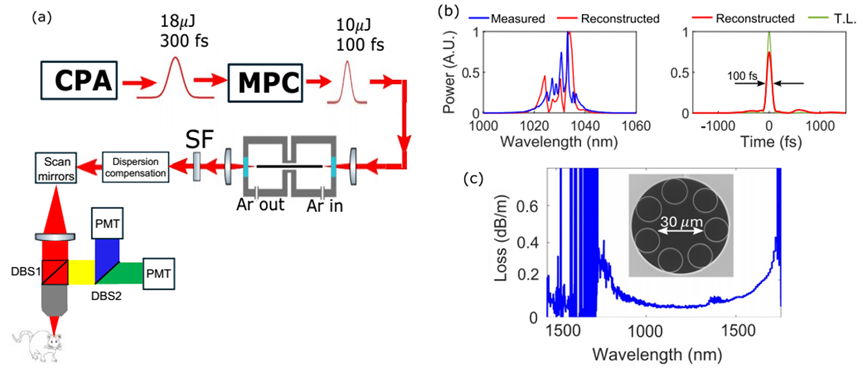
Figure 1(a) Schematic of the system setup; (b) Left: Measured and reconstructed pulse spectrum after MPC. Right: Reconstructed pulse temporal profile after MPC; (c) Transmittance of the anti-resonant fiber [1]
As depicted in Figure 2, the spectrum broadens with increasing gas pressure, culminating in a prominent component at 1300 nm. By adjusting the input pulse duration between 60 fs and 100 fs to optimize both spectral shape and temporal pulse, it was determined that 100 fs input pulses yielded the optimal results. Utilizing a long-pass filter, pulses with an energy of 960 nJ were isolated at wavelengths exceeding 1240 nm (Figure 3(a)). The pulse initially exhibited a trailing tail (Figure 3(b)), which was eliminated post-dispersion compensation, resulting in a 50 fs pulse (Figure 3(c)).

Figure 2 Output spectrum of the fiber at different gas pressures [1]
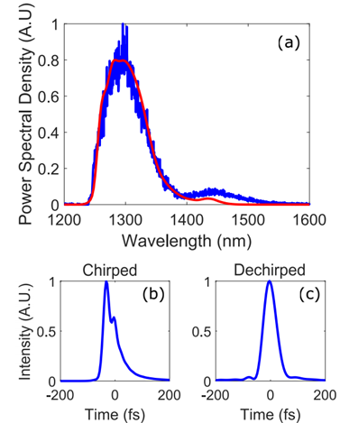
Figure 3 (a) Blue: Spectrum of the filtered band; Red: Spectrum reconstructed by the FROG algorithm for the dechirped pulse. (b) Temporal profile of the filtered band without dispersion compensation, and (c) Temporal profile of the filtered band with dispersion compensation [1]
As shown in Figure 4, with a fixed input pulse duration of 100 fs and varying gas pressures, the light source is capable of delivering megawatt-level pulses across the entire octave range from 850 nm to 1700 nm, even without dispersion compensation.
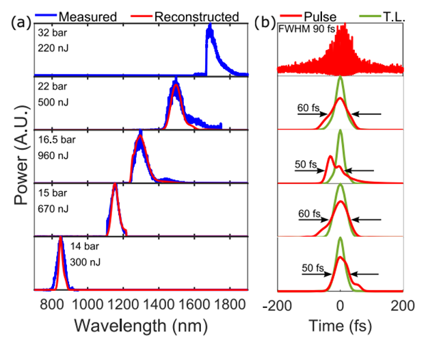
Figure 4 Filtered pulse spectra and temporal profiles from 850-1700 nm at different gas pressures [1]
Subsequently, the authors employed filtered pulses centered at 1300 nm to perform structural and functional three-photon in vivo imaging of neurons in transgenic GCaMP6s mice. Figures 5(a)-(c) showcase structural imaging of the entire cortical column of the mouse, extending down to the external capsule (depth of approximately 950 µm to 1100 µm). Spontaneous neuronal activity can be consistently detected by 3PM at depths of ~800 µm below the dura mater, with a sufficient photon count per neuron per second to enable high-fidelity calcium transient recording (Figures 5(d)-(e)).

Figure 5 In vivo three-photon imaging of the deep brain of transgenic GCaMP6s mice using a 1300 nm light source [1]
This article presents a wavelength-tunable 100 fs pulse source, leveraging the self-phase modulation effect in hollow-core fiber, capable of delivering peak powers exceeding 1 MW across the spectrum from 850 nm to 1700 nm. This innovative approach paves the way for generating high-energy ~100 fs pulses that can be broadly tuned across the most crucial wavelength windows for multiphoton microscopy, enabling deep tissue imaging. The demonstration of structural and functional three-photon fluorescence images of deep brain tissue in mice underscores the efficacy of this advanced light source.
References:
[1] Eisenberg, Y., Wang, W., Zhao, S., Hebert, E. S., Chen, Y. H., Ouzounov, D. G., ... & Wise, F. (2024). Efficient, broadly-tunable source of megawatt pulses for multiphoton microscopy based on self-phase modulation in argon-filled hollow-core fiber. arXiv preprint arXiv:2410.00889.
-
![]()
Dianping Merges into Core Local Business as Meituan Advances Towards Greater Unity
-
![]()
Reevaluating vivo's Leap into Robotics
-
![]()
iOS 18.5 In-depth Review: Does Apple's Latest Update Cater to Chinese Users?
-
![]()
Qingming Holiday Spurs Demand Surge! Will Hitchhiking Become the Next 100 Billion Travel Market?
-
![]()
Samsung S25 Claims the Title of the Price Drop King in 2025, Outpacing iPhone 16e
-
![]()
Unisound AI Races to Hong Kong IPO with Estimated Valuation of RMB 9 Billion
-
![]()
QuantGroup Garners Broad Market Acclaim, Recently Pursuing IPO
-
![]()
California Startup Unveils Groundbreaking Optical Interconnect Technology to Revolutionize AI Computing


